

Your gut does a big share of your immune work. And postbiotics may help by backing the gut lining, shaping immune signals, and making the gut less friendly to harmful microbes.
Here’s the short version: this article covers 7 postbiotic compound types and the main job each one does.
- SCFAs help fuel colon cells and support tight junctions
- Bacteriocins help limit harmful bacteria
- Microbial enzymes help lower oxidative stress
- Indole derivatives signal through AhR and support IL-22
- Peptidoglycan fragments signal through NOD1/NOD2
- Teichoic and lipoteichoic acids signal through TLR2
- EPS help support the mucus layer and steady immune tone
A few facts stand out:
- Butyrate provides about 70% of colonocyte energy
- The main SCFAs often appear in a 60:20:20 ratio: acetate, propionate, butyrate
- Some postbiotics help with barrier support
- Some act as immune cues
- Some help with microbial control
Put simply, I’d group the seven compounds into three core roles:
- Support the gut barrier
- Guide immune response
- Help keep harmful microbes in check
That matters because postbiotics are nonliving microbial compounds, so they don’t colonize the gut and don’t need to stay alive to work.
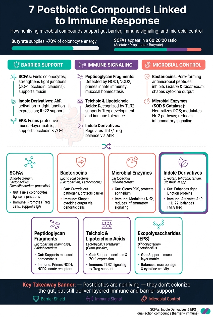
7 Postbiotic Compounds: Gut & Immune Roles at a Glance
Postbiotics Explained: The Gut Health Trend You’ll Hear More About | Suzanne Devkota
sbb-itb-1bbfe7f
Quick Comparison
| Compound | Main role | Main immune link | Main gut link |
|---|---|---|---|
| SCFAs | Cell fuel + signaling | Treg support, IgA support | Tight junctions, mucin |
| Bacteriocins | Microbe control | Cytokine signaling | Helps protect barrier from pathogens |
| Microbial enzymes | Oxidative stress control | Nrf2-linked effects | Helps limit ROS damage |
| Indole derivatives | Chemical signaling | AhR, IL-22, Treg/Th17 balance | Tight junction support |
| Peptidoglycan fragments | Immune priming | NOD1/NOD2 | Mucosal homeostasis |
| Teichoic/Lipoteichoic acids | Cell-wall signaling | TLR2, Treg support | Tight junction support |
| EPS | Surface support | Macrophage and cytokine balance | Mucus-layer support |
If you want the main takeaway fast, it’s this: different postbiotics do different jobs, and the value comes from how they work together, not from one compound alone.
How Postbiotics Affect Gut and Immune Balance
Some postbiotics - especially SCFAs - help feed colon cells and support the gut barrier.[1] One big way they do that is by increasing the expression of tight junction proteins such as zonula occludens-1 (ZO-1), occludin, and claudins.[3] These proteins help seal the spaces between epithelial cells, which limits pathogen entry.[3]
Postbiotics can also change the gut environment itself. Compounds like acetate and lactate lower intestinal pH, making the gut more acidic and less friendly to harmful bacteria.[3]
They also send signals through pattern-recognition receptors like Toll-like receptors, which helps the immune system tell commensals apart from threats. On top of that, they bind G-protein-coupled receptors on immune and epithelial cells, which helps regulate inflammation.[1][3]
Some postbiotics work at another level. Butyrate, in particular, inhibits histone deacetylases (HDACs), which promotes the development of T-regulatory (Treg) cells and supports anti-inflammatory activity.[1] In inflammatory settings marked by IL-6, that balance can shift toward Th17 responses that help clear pathogens.[3]
These pathways overlap, but each class of compound works in its own way. Next: how individual postbiotic compounds influence immunity.
1. Short-Chain Fatty Acids (SCFAs)
SCFAs are major metabolites made by gut microbes. They’re produced when helpful bacteria ferment non-digestible fibers and resistant starches in the large intestine. The three main SCFAs are acetate, propionate, and butyrate, and they’re usually present in the colon at a molar ratio of about 60:20:20. [1]
SCFAs are the best-known postbiotic mediators of gut-immune balance.
Gut-Barrier Support
Butyrate provides about 70% of the total energy colonocytes need to do their job. [5] It also helps strengthen the intestinal barrier by supporting tight-junction proteins and mucin production. SCFAs support goblet cells too, which can increase mucin and help reinforce the mucus layer.
Immune Pathway or Cell Targets
SCFAs send signals through GPR41, GPR43, and GPR109A on immune and epithelial cells. [1] Butyrate and propionate inhibit HDACs, which helps promote regulatory T cells. Acetate supports secretory IgA production by B cells. [1]
Main Producers
- Acetate: mainly from Bifidobacterium and Lactobacillus
- Propionate: mainly from Bacteroides and Phascolarctobacterium
- Butyrate: mainly from Faecalibacterium prausnitzii, Roseburia, and Eubacterium rectale
Evidence Note
Low levels of butyrate-producing bacteria, especially Faecalibacterium prausnitzii, are linked with IBD and other inflammatory conditions. [1][4]
Next up are bacterial peptides, which act more directly on competing microbes and immune signaling.
2. Bacterial Peptides and Bacteriocins
Bacteriocins are small antimicrobial peptides made by beneficial bacteria.
Primary Gut-Barrier Effect
One of their main jobs is to help protect the gut barrier by crowding out pathogens. When they inhibit enteric pathogens such as Listeria monocytogenes and Clostridium species, they can help preserve tight junction integrity and lower the risk of barrier breakdown.
Some bacteriocins do this by forming pores in pathogen cell membranes. Others interfere with cell wall synthesis. Either way, the result is simple: they make it harder for harmful microbes to gain ground in the gut. That antimicrobial action also helps shape immune signaling.
Immune Pathway or Cell Targets
Bacteriocins interact with immune-cell receptors on dendritic cells and macrophages, which helps shape cytokine output. They may also influence T-cell differentiation, supporting regulatory T cells while limiting excess Th17 activity.
Main Source or Microbial Origin
These peptides are mainly produced by lactic acid bacteria, including Lactobacillus, Bifidobacterium, and Lactococcus species.
Key Safety Note
Because bacteriocins are nonliving compounds, they cannot replicate. They're also stable during processing and storage.
Microbial enzymes work in a different way, shaping metabolism and immune signaling instead of directly suppressing microbes.
3. Microbial Enzymes
Unlike bacteriocins, these enzymes don’t work by directly holding microbes back. Instead, they help limit oxidative damage in the gut. Microbial enzymes such as superoxide dismutase (SOD) and catalase help neutralize oxidative stress, which can cut inflammation that weakens the intestinal lining.
Primary Gut-Barrier Effect
SOD and catalase help clear reactive oxygen species (ROS) before those molecules can damage the intestinal epithelium. Less oxidative stress means better support for barrier integrity.
Immune Effects
When ROS-driven stress is kept in check, inflammatory pathways in the gut lining are less likely to get switched on. These enzymes may also modulate the Nrf2 pathway, a key regulator of the antioxidant response. That can help reduce inflammatory signaling and support balanced T-cell responses.
Next: cell-wall fragments that trigger immune signaling more directly.
Main Source or Microbial Origin
SOD and catalase are mainly linked with helpful bacteria in the Lactobacillus and Bifidobacterium genera, where they are released through normal microbial activity.
4. Indole Derivatives
Indole derivatives are microbial metabolites that form when gut bacteria break down tryptophan. Unlike enzymes or antimicrobial peptides, these compounds mostly work as signaling molecules through AhR. The best-known examples are indole-3-propionic acid (IPA), indole-3-aldehyde (IAld), and indole-3-acetic acid (IAA) [5].
Primary Gut-Barrier Effect
These compounds bind to the Aryl Hydrocarbon Receptor (AhR). Once AhR turns on, it helps drive the expression of tight junction proteins like ZO-1 and occludin. That process helps the intestinal lining stay intact and supports barrier function [5].
Immune Pathway or Cell Targets
AhR activation also pushes Innate Lymphoid Cells (ILCs) to produce Interleukin-22 (IL-22), a cytokine that plays a central role in mucosal repair and the release of antimicrobial peptides [5][1]. Indole derivatives also help keep the balance between pro-inflammatory Th17 cells and anti-inflammatory regulatory T cells (Tregs), which supports immune balance [1].
Put simply, indoles act like a chemical link between what gut microbes make and how the immune system responds.
Main Source or Microbial Origin
Major producers include Lactobacillus, especially L. reuteri, along with Bifidobacterium and some Clostridium species [5][4].
Key Safety or Evidence Notes
Low AhR-ligand levels are linked to more intestinal inflammation and weaker resistance to pathogens [1].
5. Peptidoglycan Fragments
Peptidoglycan (PGN) fragments are small pieces of the bacterial cell wall released during growth, division, or lysis. In plain terms, they give the host a way to sense microbes without needing live bacteria to be present.
Primary Gut-Barrier Effect
PGN fragments may help support gut-barrier integrity by increasing tight-junction proteins such as occludin and ZO-1.
Immune Pathway or Cell Targets
PGN fragments do more than support the barrier. They also work as immune signals.
Inside cells, these fragments are detected by pattern recognition receptors (PRRs), especially NOD1 and NOD2. When they activate NOD1 and NOD2, they help shape Th17-related immune signaling and play a role in immune priming.
Main Source or Microbial Origin
PGN fragments come from both Gram-positive and Gram-negative bacteria. Common probiotic sources include Lactobacillus species, especially L. rhamnosus, and Bifidobacterium species.
Key Safety or Evidence Notes
Because PGN fragments are natural byproducts of commensal and probiotic bacteria, they usually support homeostatic immune signaling instead of driving inflammation. In a healthy gut, they tend to act more like immune cues than inflammatory triggers.
Next: teichoic and lipoteichoic acids, which also come from bacterial cell walls but signal through different pathways.
6. Teichoic and Lipoteichoic Acids
After peptidoglycan fragments, the next Gram-positive cell-wall signal is LTA. Teichoic acids are polymers found in Gram-positive cell walls, and lipoteichoic acid is the membrane-anchored form most closely linked to immune signaling.
Primary Gut-Barrier Effect
Like peptidoglycan fragments, LTA gives the immune system a nonliving cell-wall cue. But it works through TLR2, not the same route as peptidoglycan fragments. LTA from strains such as L. plantarum may help support intestinal barrier integrity by increasing the expression of tight-junction proteins, including occludin and ZO-1[5].
Immune Pathway or Cell Targets
LTA is recognized by Toll-like receptor 2 (TLR2)[5]. That signal can influence T-cell balance and may support Treg development and immune tolerance[1].
Main Source or Microbial Origin
These compounds are found in Gram-positive bacteria. Lactobacillus species, including Lactobacillus plantarum, are a prominent source[5].
7. Exopolysaccharides (EPS)
Unlike the cell-wall fragments above, EPS are secreted carbohydrates that work on the gut surface. They’re bacterial sugar polymers released into the gut environment.
Primary Gut-Barrier Effect
EPS can form a protective matrix along the gut lining. They may also help support occludin and ZO-1.
Immune Pathway or Cell Targets
EPS help maintain a stable microbiome, which in turn helps keep immune signaling balanced.
Key Safety or Evidence Notes
EPS may support barrier function and help the gut deal with everyday stress.
How These Postbiotic Compounds Work Together
These compounds don’t work in isolation. They back each other up across barrier support, immune signaling, and microbial control.
They tend to fall into three main roles. Metabolites like SCFAs and indole derivatives help strengthen the barrier and support regulatory T-cell balance. Structural fragments like peptidoglycan and teichoic acids serve as immune cues through pattern-recognition receptors. Bacteriocins and enzymes add antimicrobial pressure, which can lower stress on the barrier. EPS helps steady the mucus layer and the local inflammatory tone.
The end result is a layered defense. Each compound supports the others instead of doing the same job twice.
That layered effect helps explain why postbiotics are often used as part of broader synbiotic-postbiotic formulas.
Where a Synbiotic-Postbiotic Formula May Fit
That layered action helps explain why some routines put all three eubiotic parts into a single formula. Since these postbiotic pathways overlap, some products bring together prebiotics, probiotics, and postbiotics in one routine.
Rebiirth RE-1 is positioned as a 3-in-1 synbiotic-postbiotic formula that combines prebiotics, probiotics, and a postbiotic component. But the main idea here isn’t the brand name. It’s the approach: pairing the substrate, live strains, and postbiotic signals in one system.
Quick Reference: Postbiotic Compounds at a Glance
After the deeper profiles above, this table pulls the seven classes into one quick scan.
Use it to check each compound’s source, gut role, and immune link at a glance.
| Postbiotic Compound | Main Source | Primary Gut Effect | Immune Connection | Evidence & Safety |
|---|---|---|---|---|
| Short-Chain Fatty Acids (SCFAs) | Gut microbial fiber fermentation | Fuels colonocytes; strengthens tight junctions | Promotes Treg cell differentiation [1] | Well-studied; high safety profile |
| Bacterial Peptides and Bacteriocins | Lactic acid bacteria | Inhibits pathogenic bacteria | Supports innate immune signaling | Mainly in vitro and animal studies |
| Microbial Enzymes | Secreted by probiotic strains | Limits oxidative stress in the gut | Supports antioxidant and inflammatory balance | Well-studied for digestive support; well-tolerated |
| Indole Derivatives | Microbial tryptophan metabolism | Enhances intestinal barrier integrity | Activates AhR; regulates IL-22 [1] | Emerging human data |
| Peptidoglycan Fragments | Bacterial cell wall breakdown | Maintains mucosal homeostasis | Primes innate immunity via NOD1/NOD2 receptors | Mainly mechanistic |
| Teichoic and Lipoteichoic Acids | Gram-positive bacterial cell walls | Supports barrier signaling | Interacts with TLR2 to modulate cytokines | Extensive animal and cell studies; high stability |
| Exopolysaccharides (EPS) | Secreted by Bifidobacterium and Lactobacillus | Supports the mucus layer | Supports macrophage activity and cytokine balance | Growing clinical interest; used as safe food additives |
One useful distinction stands out: SCFAs, indole derivatives, and EPS are dual-action compounds. They help the gut lining and immune signaling at the same time [1][2].
The other groups lean a bit differently. Peptidoglycan fragments and teichoic acids are more tied to immune receptor priming. Bacteriocins and microbial enzymes, on the other hand, work more through pathogen control and oxidative balance inside the gut environment.
Because postbiotics are non-viable, they don’t replicate. That also means they’re often more stable in storage than live probiotics, which can help support more consistent, standardized dosing in supplement form.
Conclusion
Postbiotics include many microbial compounds, and they can help support gut and immune balance in different ways. Put simply, these seven compounds tend to fall into three main roles: barrier support, immune signaling, and microbial control.
Some, like peptidoglycan fragments and teichoic acids, work as structural signals that help prime immune receptors. Others, like SCFAs and indole derivatives, act more like chemical messengers, shifting immune-cell activity. And they do this without colonizing the gut or needing to stay viable.
The key point is simple: different postbiotic classes do different jobs in the immune system. So if you want to support the back-and-forth communication behind immune balance, a formula that includes more than one class may make more sense.
That layered model helps explain why a combined formula can be a good fit. Rebiirth RE-1 follows this approach with a 3-in-1 synbiotic-postbiotic formula.
FAQs
What are postbiotics, exactly?
Postbiotics are non-living microbial parts and byproducts made during fermentation. They include SCFAs, enzymes, peptides, and cell wall fragments.
Unlike live probiotics, postbiotics work more directly. They can help strengthen the gut lining, neutralize harmful substances, and support immune response and inflammation control.
How are postbiotics different from probiotics?
Probiotics are live microorganisms that help support gut balance. Postbiotics are the non-living byproducts and metabolites those microbes make during fermentation.
Put simply, probiotics are the microbes themselves. Postbiotics are the compounds they leave behind - like short-chain fatty acids, enzymes, and peptides - that do much of the work when it comes to health support.
How can I support postbiotic production in my gut?
Support postbiotic production by helping good gut bacteria do their job. Because postbiotics are created during fermentation, the aim is to keep your microbiome active and in balance.
A few foods and products can help:
- Eat prebiotic-rich foods like garlic, onions, leeks, asparagus, and artichokes
- Include resistant starches, such as green banana powder
- Add fermented foods like yogurt, kefir, sauerkraut, kimchi, and miso
- Consider Rebiirth RE-1, which combines prebiotics, probiotics, and postbiotics