

Your gut helps train your immune system every day. In fact, about 70% to 80% of immune cells are linked to the gut, so when gut microbes shift, immune balance can shift too.
Here’s the short version:
- The gut lining controls contact between microbes and immune cells.
- GALT - including Peyer’s patches and mesenteric lymph nodes - sorts signals into tolerance, IgA output, or inflammation.
- Innate cells react first through TLRs, NLRs, and cytokines.
- Adaptive cells like Tregs, Th17 cells, and B cells shape longer-term immune patterns.
- Microbial metabolites such as butyrate, acetate, and propionate can affect immune activity inside and outside the gut.
- When the microbiome stays balanced, the barrier tends to stay tight. When dysbiosis sets in, LPS can move into the blood and drive inflammation.
If I had to boil the whole article down to one idea, it’s this: gut health and immune control are tied together through barrier function, immune training, and microbe-made signals. That link helps explain why shifts in the microbiome are connected with allergy, inflammation, and other gut-immune problems.
A few facts stand out:
- sIgA coats microbes and helps keep them in the gut lumen.
- SCFAs made from fiber fermentation can push immune cells toward a calmer state.
- Tregs help limit immune overreaction, while Th17 cells help fight threats but can add to tissue damage if control is lost.
- Secondary bile acids also shape immune-cell behavior through receptors such as TGR5 and FXR.
Quick view of the gut-immune pathway:
| Step | What happens | Main players |
|---|---|---|
| 1 | Microbes meet the gut barrier | Mucus, epithelial cells, tight junctions, sIgA |
| 2 | Signals get sampled | Dendritic cells, M cells, microbial metabolites |
| 3 | Immune decisions are made | Peyer’s patches, MLNs, macrophages, B cells, T cells |
| 4 | Cells act | Tregs, Th17 cells, IgA-producing B cells, ILC3s |
| 5 | Effects spread past the gut | SCFAs, bile acids, blood-borne inflammatory signals |
So if you want to understand how gut microbes shape immunity, the answer is pretty simple: they do it through constant two-way signaling at the barrier, in gut immune tissue, and through metabolites that can move through the body.
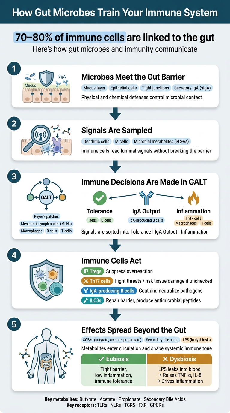
How Gut Microbes Train Your Immune System: A 5-Step Pathway
How Does Your Gut Microbiota Impact Your Immune System | Modern Immunoinformatics
sbb-itb-1bbfe7f
The Gut Barrier: Where Microbes First Meet the Immune System
At the gut lining, microbes run into the body's first line of defense: mucus, epithelial cells, and local immune defenses. Each layer helps control how much microbial contact reaches immune sensors.
Mucus, Epithelial Cells, and Tight Junctions
Goblet cells secrete mucus, and that mucus keeps most microbes away from the epithelial surface. In plain terms, it acts like a buffer zone. It also helps control which microbial products make it through to immune sensors.
Right behind that mucus layer are the intestinal epithelial cells (IECs). These cells don't just sit there as a wall. They also work as a signaling interface, using PRRs, including TLRs, to detect microbial patterns such as LPS and flagellin. TLR5 sits on the basolateral side, which helps limit unnecessary responses to commensals.
The epithelial cells are held tightly together by tight junctions. These protein complexes seal the spaces between cells and regulate what can pass through.
Taken together, these defenses don't shut down all microbial contact. They manage how much of it gets through to immune cells.
Secretory IgA and Local Containment
Another major part of the gut barrier is secretory IgA (sIgA). This antibody is produced locally and released into the gut lumen, where it coats microbes. When sIgA binds to bacteria, it limits how well they stick to the epithelial surface and how far they move into tissue. The result is local containment rather than constant escalation.
When that containment isn't enough, the immune system still has ways to check what's happening in the lumen.
From Lumen to Lamina Propria: How Signals Are Transferred
Even with the barrier in place, the immune system still needs a steady read on the gut lumen. Two specialized mechanisms help make that happen without tearing down the barrier.
One involves dendritic cells (DCs). These cells extend projections between epithelial cells to sample luminal bacteria without breaking tight junctions.
The other involves microbial metabolites, especially short-chain fatty acids (SCFAs) made when gut bacteria ferment dietary fiber. SCFAs are one route by which gut microbes affect immune activity beyond the barrier. They enter the lamina propria and alter immune-cell behavior and gene regulation.
So the lamina propria isn't working blind. It gets a steady stream of information from the lumen, which helps immune cells decide whether to tolerate or respond.
These barrier-level signals then feed into GALT, where immune cells decide whether to tolerate or respond. From there, the same signals move into GALT for immune training and decision-making.
| Mechanism | Structure Involved | Role |
|---|---|---|
| Physical separation | Mucus layer | Keeps most microbes away from epithelial tissue |
| Microbial signal detection | Epithelial TLRs | Reads bacterial patterns and helps shape immune responses |
| Paracellular control | Tight junctions | Regulates what passes between epithelial cells |
| Immune containment | Secretory IgA | Coats microbes to limit tissue penetration |
| Direct lumen sampling | Dendritic cells | Samples bacteria without disrupting barrier integrity |
| Metabolite signaling | SCFAs | Enter the lamina propria and alter immune-cell behavior and gene regulation |
How Gut-Associated Lymphoid Tissue Turns Microbial Signals Into Immune Decisions
Once microbial antigens and metabolites get past the gut barrier, they move into a connected set of immune structures called gut-associated lymphoid tissue (GALT). This is where the immune system starts making a key call: ignore and tolerate, or gear up and defend. In practice, Peyer's patches and mesenteric lymph nodes (MLNs) help turn those microbial inputs into T- and B-cell responses.
Peyer's Patches, M Cells, and Antigen Sampling
Peyer's patches are organized lymphoid tissues in the small intestine. They act as main sites for sampling antigens from the gut lumen. Their follicle-associated epithelium contains specialized microfold cells, or M cells, which transport luminal antigens to immune cells beneath the epithelial layer.
Once material moves through an M cell, it reaches dendritic cells and macrophages located below the epithelium. These cells detect microbial patterns such as LPS and flagellin through pattern-recognition receptors. That step helps connect early innate sensing with later T- and B-cell responses.
Beyond Peyer's patches, the small intestine also contains isolated lymphoid follicles (ILFs), which are smaller immune structures found on the antimesenteric wall and take part in mucosal immune surveillance [1]. Put simply, ILFs add another checkpoint in the gut's watch system.
Those early signals don't stay local for long. They are further shaped in the mesenteric lymph nodes.
Mesenteric Lymph Nodes and Immune Education
After sampling, dendritic cells and macrophages carry microbial information to the mesenteric lymph nodes (MLNs), which drain the gut. In the MLNs, antigen-presenting cells guide T- and B-cell responses toward tolerance, IgA production, or active defense.
This is where the immune system starts sorting friend from foe. Some signals lead to restraint. Others push the system toward protection.
Key Resident Cells in GALT
GALT contains several immune cell types that work together to keep mucosal responses in check.
| Cell Type | Primary Role in GALT |
|---|---|
| Dendritic cells | Sample antigens, present them to T and B cells, and direct tolerance or defense |
| Macrophages | Phagocytose pathogens, produce cytokines, and support antigen presentation |
| B cells | Differentiate into IgA-secreting plasma cells after microbial stimulation |
| T regulatory cells (Tregs) | Suppress excessive inflammation and promote tolerance to commensals |
| Th17 cells | Drive inflammatory defense against pathogens; overactivation is linked to disease |
Microbial signals also push B cells to produce IgA. That IgA is then secreted back into the gut lumen, where it coats mucosal surfaces and helps block harmful bacterial invasion [1]. It's a tight feedback loop between the lumen, GALT, and the gut barrier, and that loop helps keep mucosal immunity in balance.
These GALT-level decisions set the stage for the innate sensing, T-cell balance, and IgA responses that come next.
Innate and Adaptive Immune Cells That Respond to Gut Microbes
Inside GALT, microbial signals are turned into two layers of immune response: a fast innate response and a slower adaptive one.
Innate Sensing Through TLRs, NOD-Like Receptors, and Cytokines
Innate immune cells move first. Dendritic cells, macrophages, neutrophils, and ILC3s carry PRRs such as TLRs and NLRs. These receptors detect microbial patterns like lipopolysaccharide (LPS) and flagellin.
What happens next depends on the setting. Dendritic cells can make IL-10 to support tolerance or IL-12 to push inflammation [1]. Macrophages that sense LPS through TLR4 can release TNF-α and IL-6. By contrast, signals from commensal bacteria through TLR2 can support IL-10 production and a more anti-inflammatory response [2].
Epithelial cells are part of this first line too. They express TLRs and respond by releasing antimicrobial peptides, increasing mucus secretion, and strengthening the physical barrier. ILC3s react to microbial metabolites and cytokines in ways that help repair the barrier and support antimicrobial peptide production.
| Cell Type | Sensing Route | Immune Outcome |
|---|---|---|
| Dendritic Cells | TLRs, NLRs | IL-10 or IL-12 |
| Macrophages | TLR4 or TLR2 | TNF-α, IL-6, or IL-10 |
| Epithelial Cells | Apical/basolateral TLRs | Barrier repair; antimicrobial peptide release; mucus secretion |
| Neutrophils | PRRs, chemokine receptors | Rapid recruitment; antimicrobial activity; acute inflammation |
| ILC3s | Microbial metabolites and cytokines | Promote barrier repair and antimicrobial peptides |
These cytokines don’t act in isolation. They help decide what T cells and B cells do next.
How Gut Microbes Shape Tregs, Th17 Cells, and IgA-Producing B Cells
Innate signals then guide Tregs, Th17 cells, and IgA-producing B cells.
Tregs help prevent too much inflammation. Commensal bacteria and probiotics, including Bifidobacterium, support Treg differentiation. These cells then secrete IL-10 and TGF-β to calm overactive immune responses [1][2]. You can think of them as part of the gut’s braking system.
Th17 cells play a key role in mucosal defense and can be driven by bacteria like Segmented Filamentous Bacteria (SFB). But there’s a trade-off. If Tregs don’t keep them under control, Th17 cells can add to inflammation and tissue damage.
IgA-producing B cells are another major part of mucosal defense. Secretory IgA coats and neutralizes harmful bacteria before they invade the mucosal surface.
| Cell Type | Main Microbial Trigger | Function in Mucosal Immunity |
|---|---|---|
| Tregs | Commensal metabolites (SCFAs), IL-10 | Maintains tolerance; suppresses autoimmunity and chronic inflammation |
| Th17 Cells | Segmented Filamentous Bacteria (SFB) | Drives mucosal defense; can cause inflammation if unchecked |
| IgA-Producing B Cells | Commensal bacteria stimulation in GALT | Produces sIgA to neutralize pathogens and maintain barrier integrity |
When dysbiosis shifts this balance, inflammatory signals can move past the gut lining. Those cell-level choices also shape how far immune effects spread beyond the gut.
Microbial Metabolites, Systemic Signaling, and Why Microbiome Balance Matters
Short-Chain Fatty Acids, Bile Acids, and Epigenetic Signaling
Tregs, Th17 cells, and IgA-producing B cells don't act in isolation. They're shaped, in part, by microbial metabolites made in the gut.
Among the best-studied are short-chain fatty acids (SCFAs): acetate, propionate, and butyrate. Commensal bacteria such as Bifidobacterium and Lactobacillus make these compounds when they ferment prebiotic fibers like inulin and GOS. From there, SCFAs signal through G-protein coupled receptors and also act as HDAC inhibitors. Put simply, they can influence gene activity in immune cells and steer them toward a more tolerant, anti-inflammatory state.
Secondary bile acids work through a different pathway. Gut bacteria convert primary bile acids made by the liver into secondary forms that move through the enterohepatic system. These metabolites interact with receptors such as TGR5 and FXR, affecting T-cell balance and metabolic signaling beyond the gut.
| Metabolite Class | Key Examples | Primary Source | Signaling Route | Immune Effect |
|---|---|---|---|---|
| Short-Chain Fatty Acids | Butyrate, Acetate, Propionate | Fiber fermentation by commensals | GPCRs; HDAC inhibition; NF-κB/PPARα inhibition | Supports Tregs; reduces inflammation |
| Secondary Bile Acids | Deoxycholic acid, Lithocholic acid | Microbial transformation of primary bile acids | Enterohepatic circulation; TGR5 and FXR receptors | Modulates T-cell balance; influences metabolic signaling |
These signals matter most once they cross the gut barrier and start shaping immune activity in the rest of the body.
How Gut Signals Reach the Blood and Peripheral Immune System
After metabolites cross the intestinal barrier, they can affect immune tone in the blood and in distant tissues. Butyrate is a good example. It can enter circulation and help calm inflammatory signaling away from the gut.
That barrier is the gatekeeper. When it stays intact, metabolites can send body-wide signals without setting off inflammation. When it breaks down, the story changes. LPS can slip into circulation and drive inflammatory signaling instead.
Eubiosis, Dysbiosis, and Microbiome Restoration
When these gut signals change, immune balance changes with them.
In a balanced microbiome state, called eubiosis, commensal bacteria help support SCFA production, IgA secretion, and a low inflammatory tone [1]. The gut barrier stays tight, inflammation stays in check, and the immune system is better able to tell the difference between a threat and a harmless signal.
Dysbiosis turns that pattern on its head. A shift toward pathogenic species such as E. coli or Salmonella can lower SCFA production and thin the mucus barrier. That can let LPS leak into circulation, which raises TNF-α and IL-8 and pushes T-helper balance toward inflammatory and allergic disease [1].
This is why microbiome restoration gets so much attention. Synbiotic approaches have shown the most promise here. Rebirth RE-1™ is a 3-in-1 eubiotic synbiotic designed to support microbiome balance through prebiotics, probiotics, and postbiotics.
FAQs
How does the gut barrier prevent immune overreaction?
The gut barrier helps stop the immune system from going into overdrive. It acts like a selective physical and biochemical filter between the body and possible pathogens.
Tight junction proteins seal intestinal cells together. On top of that, a protective mucus layer traps harmful molecules. Together, these defenses limit toxins from getting into the bloodstream and help the immune system tell the difference between harmless substances and actual threats.
What happens to immunity when gut bacteria become imbalanced?
When gut bacteria get out of balance, a state called dysbiosis, the immune system doesn’t work as well as it should.
Here’s what starts to go wrong: harmful bacteria can weaken the gut barrier, lower helpful metabolites such as short-chain fatty acids, and throw off the balance between Th17 cells and regulatory T cells.
That shift can weaken mucosal defenses, fuel chronic inflammation, and increase the risk of autoimmune disorders, inflammatory bowel disease, and infections.
How do gut-made metabolites affect immune cells outside the gut?
Gut-made metabolites, especially short-chain fatty acids like acetate, propionate, and butyrate, do more than work inside the digestive tract. Once they enter the bloodstream, they can influence immune activity throughout the body by signaling to immune cells through receptors or by changing gene expression.
That matters because these compounds can shape cytokine production, support anti-inflammatory regulatory T cells, and help lower systemic inflammation. Rebiirth RE-1™ supports this process by helping restore microbiome balance and improve production of these metabolites.